KPSS Phosphosite Profiling Services
Kinetworks™
Phosphosite Screen KPSS 1.3 Profiling
Kinetworks™
Phosphosite Screen KPSS 10.1 Profiling
Kinetworks™
Phosphosite Screen KPSS 11.0 Profiling
Kinetworks™
Phosphosite Screen KPSS 12.1 Profiling
The Kinetworks™ KPSS phosphosite profiling services permits true functional proteomics analyses. Phenotypic changes in cells correlate best with the active forms of proteins, which are tightly regulated by reversible protein phosphorylation. In humans, at least 538 protein kinases regulate cellular functions by catalyzing the phosphorylation of most of the proteome. The human phosphoproteome may well contain over a million different phosphosites. Kinexus utilizes 4 standard KPSS screens as well as customized services to track over 333 unique and known phosphosites. Due to cross-reactivities, several hundred additional phosphosites may be detected. While this is only a small representative sampling of the existing phosphosites, our Kinetworks™ KPSS phosphosite profiling services are still unmatched for breadth, sensitivity, quantification, reproducibility, speed of turnaround, and cost.
The Kinetworks™ KPSS screens utilize the best of over 800 phosphosite antibodies that have been provided by six leading suppliers and independently tested by Kinexus. Each Kinetworks™ KPSS screen tracks from 37 to 44 known phosphorylation sites with 500 µg of cell/tissue lysate protein. The KPSS screens have been developed to work with human, rat and mouse samples, but due to the high conservation of phosphorylation sites in evolution, they have very broad utility. For example, nearly half of the human phosphorylation sites tracked in the KPSS screens are also successfully detected in frog oocytes.
The various Kinetworks™ KPSS screens are organized along thematic lines: KPSS 1.3 provides broad coverage of signalling pathways; KPSS 10.1 tracks cell cycle status; KPSS 11.0 tracks a wide range of protein kinases (except those covered in the KPSS 10.0); and KPSS 12.1 monitors the substrates of protein kinases.
Our Kinetworks™ KPSS services also serve as highly effective means for the discovery of novel phosphorylation sites in proteins. In addition to the target phosphoproteins, the KPSS screens monitor at least another 300 unknown phosphoproteins, which are detected by cross-reactivity with our panels of anti-phosphosite antibodies. Using an anti-phosphosite antibody that cross-reacts with an unknown phosphoprotein, it is feasible to establish the identity of that phosphoprotein by mass spectrometry and quickly locate the position of the phosphorylation site within the phosphoprotein by epitope alignment. Clients that are interested in identifying cross-reactive proteins from Kinetworks™ immunoblots should contact our technical representatives for a quotation for this custom service. Moreover, with antibody-based phosphosite discovery, it is possible to rapidly follow up with Western blotting, immunoprecipitation, immunohistochemistry and flow cytometry studies.
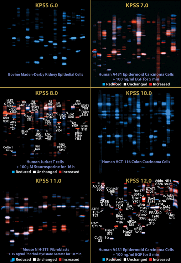
Due to the formulation of the Kinetworks™ multi-immunoblots and the running conditions for electrophoresis, it is sometimes possible to detect phosphorylation-dependent band shifts in proteins. Therefore, the KPSS screens can visualize changes in the phosphorylation of proteins at sites that are distinct from those targeted by the anti-phosphosite antibodies. In the accompanying animation and colored immunoblot overlays provided below, there are many examples of band-shifted phosphoproteins with diverse treatments of cells. Kinexus provides presentation and publication-ready figures of Kinetworks™ colored immunoblots overlays as an added service (see our Graphics Services).
Due to the formulation of the Kinetworks™ multi-immunoblots and the running conditions for electrophoresis, it is sometimes possible to detect phosphorylation-dependent band shifts in proteins. Therefore, the KPSS screens can visualize changes in the phosphorylation of proteins at sites that are distinct from those targeted by the anti-phosphosite antibodies. In the accompanying animation and colored immunoblot overlays provided below, there are many examples of band-shifted phosphoproteins with diverse treatments of cells. Kinexus provides presentation and publication-ready figures of Kinetworks™ colored immunoblots overlays as an added service (see our Graphics Services).

Download 1 page profile sheet about the Kinetworks™ KPSS Screens Services


