Kinex™ Antibody Microarrays
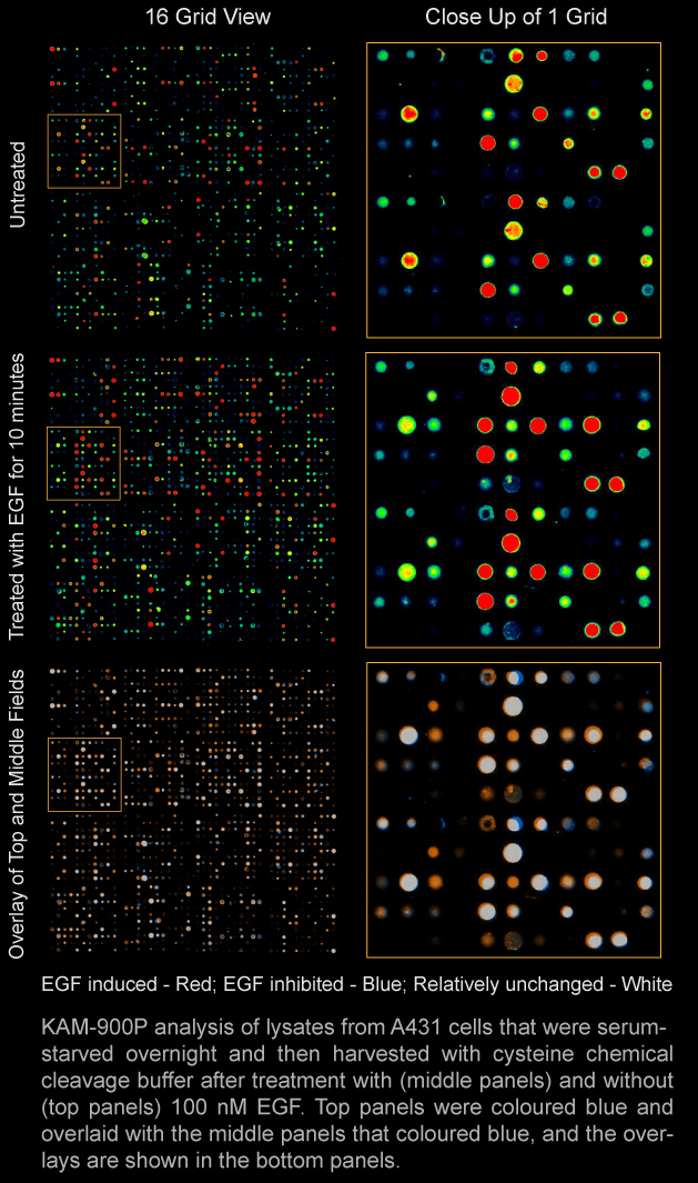
The Kinex™ antibody microarray (KAM) services provide a convenient and extremely cost-effective means for the discovery of productive research leads such as biomarkers and insights into signal transduction protein regulation. These services utilize our antibody microarrays to track the differential binding of dye- or biotin-labelled proteins in lysates prepared from cells and tissues. The results can yield useful insights into differences in protein expression, covalent modifications such as phosphorylation, and protein-protein interactions, and define antibody reagents that can be used to follow up on these findings.
Kinex™ KAM antibody microarrays permit the simultaneous probing of hundreds of target proteins and phosphorylation sites with as little as 25 µg of crude cell or tissue lysate protein. No other proteomics technology, including mass spectrometry, can compete with antibody microarrays for the directed study of target proteins in lysate samples in terms of sensitivity, speed, reproducibility, dynamic range, and cost. Therefore, the antibody microarray is a particularly attractive initial route for taking a systems biology, proteomics approach to studying human diseases and experimental model systems.
Presently, Kinexus offers the KAM-1325 antibody microarray in its services and as a stand-alone product in kits. The KAM-1325 chip features about 875 phosphosite-specific antibodies and 451 pan-specific antibodies for many of the same phosphoprotein targets. Approximately 627 unique proteins are tracked. Each antibody is printed twice in two separate fields, which allows for duplicate measurements for each of two lysate samples separately on the two halves of the same chip. About 53% of the phosphosite-specific antibodies target regulatory phosphorylation sites on over 170 different protein kinases, and 28.5% of all of the antibodies are pan-specific antibodies that monitor the expression levels of these same protein kinases. In terms of unique target proteins, these include 255 protein-serine/threonine kinases (including 12 dual-specificity kinases), 76 protein-tyrosine kinases, 104 transcription factors; 26 protein and lipid phosphatases, 49 other metabolic enzymes, 17 adaptor/scaffold proteins, and 149 other target proteins. Presently, we offer services with this microarray that permit detections of changes in protein expression levels and specific phosphosites (KAM-1325) and protein-tyrosine phosphorylation (KAM-1325-pY). Clients should contact us regarding alternative detection formats for other types of covalent modification and protein- and drug-interactions.
Our KAM-1150 chip features about 1150 different pan-specific antibodies that provide coverage for over 700 distinct signalling proteins. Approximately 581 antibodies target 304 different protein kinases, 87 antibodies target 45 protein phosphatases, and 49 antibodies target 20 stress proteins. Other major thematic families of target proteins include transcription factors, adapter/scaffold proteins; cell cycle; apoptosis, oncoproteins and ion channels. With the KAM-1150 microarray, we have developed a range of alternative detection techniques to track protein expressions, covalent modifications, protein-protein and protein-drug interactions. Presently, we offer services with this microarray that permit detections of changes in protein expression levels (KAM-1150E) and protein-tyrosine phosphorylation (KAM-1150PY). Clients should contact us regarding alternative detection formats for other types of covalent modification and protein- and drug-interactions with this microarray.
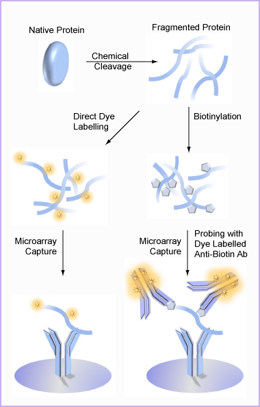
With our Kinex™ antibody microarray services, both cell/tissue lysate samples are directly labeled with the same dye mixture or biotinylated and analyzed separately on the same Kinex™ microarray chip. These approaches avoid the misleading results that can arise from the differential binding of different dyes to proteins that is problematic with the two dye competitive system used with other commercial antibody microarrays and the DIGE 2D gel electrophoresis technique.
Non-denatured proteins are typically analyzed on commercial antibody microarrays offered by other vendors. This option is also available with the Kinex™ KAM antibody microarrays. However, there is increased opportunity for false positives and false negatives due to antibody cross-reactivity, protein-protein interactions, and blocked epitopes in protein complexes. From our internal studies with cells from different species, only 30 to 45% of the protein changes detected on Kinex™ KAM antibody microarrays were reproduced by immunoblotting.
To reduce the rate of false positives and negatives from the problem of protein-protein interactions, we have added the option of having the lysate proteins subjected to chemical cleavage in a manner that preserves the integrity of most protein phosphorylation sites, especially when this is performed at the time of chemical cleavage. It should be noted that even with chemical cleavage, some phospho-epitopes may not be accessible due to internal interactions with flanking arginine and lysine residues that can mask the phosphosite from phosphosite antibodies. This issue is evident, for example with the “TEY” dual phosphosites in the ERK1 and ERK2 MAP kinases. With the chemical cleavage step, lysate samples are highly stable at ambient temperatures, and it is feasible to courier lysate samples to Kinexus for KAM analyses without the need for refrigeration or freezing during shipping.
Apart from the issues of false positives and false negatives due to unforeseen antibody cross-reactivities and protein-protein interactions, with the high sensitivity of antibody microarray detection, about 20 to 30% of the time, the target proteins are not easily visualized by immunoblotting in the tested lysates. This is despite strong detection with the same antibodies on our microarrays. Therefore, we highly recommend that any interesting Kinex™ results that clients may wish to follow up should be confirmed next by Western blotting. Unlike other vendors that sell antibody microarrays, we offer a cost-effective, custom Western blotting service (Kinetworks™ KCPS 1.0) that permits up to 18 antibodies from our Kinex™ antibody microarrays to be used at a time for such validation studies. Our Custom Kinetworks™ KCSS 1.0 service also allows clients to choose any 3 target proteins (of different molecular weight) to be quantified in 8 different samples side-by-side on the same immunoblot. The availability of these Kinetworks™ analyses is an important distinguishing feature of our antibody microarray services as clients can have their research leads conveniently and inexpensively confirmed.
Another unique feature of our Kinex™ KAM Antibody Microarray Services is that clients can freely view our extensive databases of Kinex™ Antibody Microarray and Kinetworks™ Multi-immunoblotting results that have been generated from nearly a decade of service provision by Kinexus. Our KiNET-AM database has the data from the analysis of over 4000 cell and tissue lysates on our antibody microarrays. Open access queries of the KiNET Databank can be performed based on protein, model system and treatment searches. This ability for our customers to compare their own Kinex™ Antibody Microarray results with thousands of other studies undertaken with the same methodology, reagents and equipment is not available from any other vendors for microarray products or services.
Furthermore, as part of the KAM-1325 reports provided to our clients, signalling pathway analyses with our Kinection Pathway Maps is also available. Over 220 Kinections pathway maps are linked with each report, and these maps are also freely downloadable from our PhosphoNET and KinaseNET websites. We also offer our KiNetscape Mapping Service to connect those proteins that demonstrate the greatest alterations in expression or phosphorylation into network maps that provide either qualitative or quantitative representations of the changes.
Our Kinex™ services provides researchers with access to antibody microarray technology for application to their research programs without the need for special expertise and expensive equipment such as microarray scanners and quantification software. Our goal is to keep the costs for our Kinex™ services lower than if clients purchased the microarrays and tried to perform the analyses in their own laboratory. Clients can send their cell/tissue lysates by courier to Kinexus. For an extra fee, we can also prepared lysates from frozen tissue or cell pellets. We perform the dye-labelling of the extracts, the incubations with the antibody microarrays, scanning and quantitation of the antibody spots, and the preparation and delivery of a summary report back to client within 4 weeks. Alternatively, clients can opt to utilize our Kinex™ KAM-1325 Antibody Microarray Kit. Users of this product may also send their processed KAM-1325 chips back to Kinexus for free scanning and if desired, preparation of a KAM-1325 Report for an additional fee. Instructional videos are provided with the Antibody Microarray Kit as well as available for viewing on the Kinexus Bioinformatics You-Tube Channel.


Download editable versions of the Customer Information Package forms.

