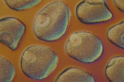
Pisaster ochraceus seastar oocytes undergoing germinal vesicle breakdown in response to treatment with 1-methyladenine. Seastar oocytes typically have a diameter of about 150 microns and are shown under phase contrast to visualize the nucleus and nucleolus. A photograph was taken every minute for a period of 30 minutes to 90 minutes after the addition of the 1-methyladenine. During this period, there is a large burst in protein phosphorylation. Kinexus has performed detailed analysis of this model system in order to further knowledge about cell cycle regulation by protein kinases. (Credits: Gordon Leung and Harry Paddon)