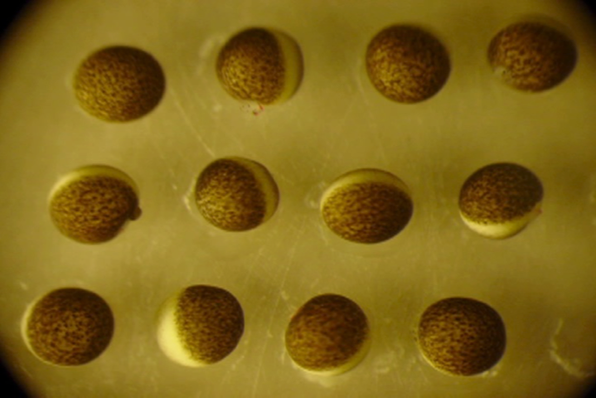
Xenopus laevis African clawed frog oocytes undergoing germinal vesicle breakdown in response to treatment with progesterone. Frog oocytes typically have a diameter of about 1.50 millimeters and are shown under normal light to visualize the release of yolk proteins at the top of the dark animal hemisphere of these oocytes following the disintegration of the nuclear membrane. A photograph was taken every 10 minutes for a period of 4 h to 9 hours after the addition of the progesterone. During this period, there is a large burst in protein phosphorylation. Kinexus has performed detailed analysis of this model system in order to further knowledge about cell cycle regulation by protein kinases. (Credits: Gordon Leung and Harry Paddon)