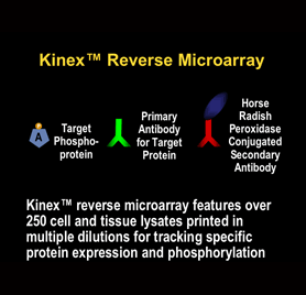
KINEX™ Reverse Lysate Microarrays
The vast majority of the proteins encoded by the genomes of humans and other species remain poorly characterized with respect to their distribution, functions and regulation. Our Kinex™ Reverse Lysate Microarray services uniquely permit the rapid simultaneous quantification of specific proteins for their distribution and regulation from analysis of hundreds of lysates from diverse cells and tissues.This is ideal for biomarker validation studies. With this convenient, cost effective and powerful service, clients can have over 230 cell and tissue lysates probed by Kinexus with one of hundreds of antibodies of their choice. Our Standard service permits the characterization of cell signalling proteins for their regulation in response to a wide range of drugs and hormones in 12 diverse human tissue cell lines, in maturing frog oocytes, and their distribution in monkey, mouse and rat tissues. These lysates are printed in triplicate at four dilutions on the Standard Kinex™ Reverse Lysate Microarray to ensure linearity of detection when the chip is probed with an antibody that specifically recognizes the target protein. Deliverables include a TIFF image of the scanned antibody probed microarray and an Excel spreadsheet with quantification of all of the lysate spots on the microarray. For the drug and hormonal treatments, the tumour cell lysates have been generated from dose response and time course studies that have been performed in-house by Kinexus. Clients may choose the probing antibody for use with the Kinex™ Reverse Microarray from our panel of over 800 available antibodies or have Kinexus perform the analysis with antibodies that they supply. This service is particularly ideal for the characterization of novel proteins that are identified initially with our Kinex™ Antibody Microarray service and have been subsequently shown with our Kinetworks™ Custom services to be specific for immune detection.
We now offer a Custom Kinex™ Reverse Lysate Microarray Service in which we can print custom chips with about 250 different cell or tissue lysates that are provided by our clients for analysis.
We now offer a Custom Kinex™ Reverse Lysate Microarray Service in which we can print custom chips with about 250 different cell or tissue lysates that are provided by our clients for analysis.
At least 20 chips must be ordered to take advantage of this service. These custom lysate microarray can be probed with antibodies available from Kinexus or those that are supplied by our clients for this service.
As the specificity of immunoreactivity of antibody can vary depending on the cell or tissue extract under examination, it may be desirable to confirm key results by immunoblotting. Any interesting Kinex™ results that clients may wish to follow up can be validated by Western blotting with the same lysates used on the Kinex™ Reverse Lysate Microarray. Our Custom Kinetworks™ KCSS 1.0 service allows clients to choose any 3 target proteins (of different molecular weight) to be quantified in 8 different samples side-by-side on the same immunoblot. The cell and tissue lysate samples from our In Vivo Services, which are used on the Kinex™ Reverse Lysate Microarray, may be used at no extra cost with the Custom Kinetworks™ KCSS 1.0 service. The availability of Kinetworks™ analyses is an important distinguishing feature of our Kinex™ microarray services as clients can have their research leads conveniently and inexpensively confirmed. Further information about the expression or phosphorylation of leads can be obtained through query of our KiNET™ on-line databank with results from over 6000 Kinetworks™ immunoblots.
A notable advantage of the Kinex™ Reverse Lysate Microarray approach as compared to studies with tissue microarrays is the better retention of soluble cytoplasmic proteins in the reverse lysate microarrays. Kinexus has determined that about three quarters of most cell signalling proteins are found in the cytosol of cells, which is typically lost in fixed cell preparations, or stain in a very diffuse pattern and are difficult to quantify for expression. Consequently, changes in protein expression or phosphorylation in response to a cellular perturbation is more accurately represented with the Kinex™ Reverse Lysate Microarray.
The following is a brief listing of the lysates from human tumour cell lines and tissues from other species that are currently available on the Kinex™ Reverse Lysate Microarray. The precise content on the Kinex™ Reverse Lysate Microarray is subject to change as we continually aim to improve and expand the range of cell and tissue lysates that it features.
A431
Skin epidermoid carcinoma cells – treated with epidermal growth factor
A549
Lung carcinoma cells – treated with interferon-gamma
HCT116
Colon carcinoma cells – treated with nocozadole
HEK 293
Female fetal kidney cells – treated with anisomycin
HeLa
Cervix epithelial adenocarcinoma cells – treated with tumour necrosis factor-alpha
HepG2
Liver carcinoma cells – treated with insulin
HL-60
Peripheral blood promyeloblasts – treated with staurosporine
HUV-EC
Umbilical vein endothelial cells – treated with phorbol ester
Jurkat
T cell leukemia cells – treated with phorbol ester
MCF-7
Breast epithelial adenocarcinoma cells – treated with insulin
PC-3
Prostate adenocarcinoma cells – treated with taxol
T98G
Brain glioblastoma cells – treated with platelet-derived growth factor
Xenopus frog oocytes – treated with progesterone
Rhesus male monkey organs and tissues
Mouse male and female organs and tissues
Rat male and female organs and tissues


Download Kinex™ Reverse Lysate Microarray Services Fillable Forms in .doc format.


